Protein Structure answers37
Protein Structure - Answers
Question 1. Look at the method X-RAY diffraction (Å). Which of the complexes is of better quality and why?
The PDB complex 7SBD, has the best resolution (lower Å).
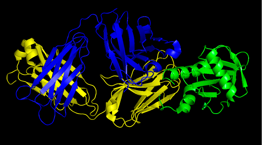
Question 2. Provide a screenshot of the complex.
Commands:
fetch 7SBD remove hetatm util.color_chains("(all)",_self=cmd) color blue, chain H color yellow, chain L select antibody, chain H+L cmd.show("surface" ,"antibody")
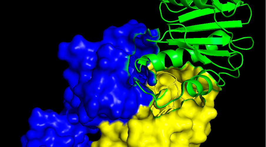
Question 3. Provide a screenshot of the interaction surface. Which secondary structures of the allergen is the antibody interacting with: alpha helices, beta-sheets or loops?
In the allergen side is mostly alpha helices and loops. If you want to analyse the antibody surface too (which is called paratope) we might need to go back to the cartoon view again. There are three well known loops called CDR1, CDR2 and CDR3, both in the light (L) chain and in the heavy (H) chain.

